anatomy and physiology of respiratory system includes…
Table of Contents
anatomy and physiology of respiratory system
The thoracic cage consists of 12 thoracic vertebrae, 12 ribs, the sternum, and costal cartilage.
The respiratory passages, consist of the upper airways1, including the nose, pharynx, and larynx;
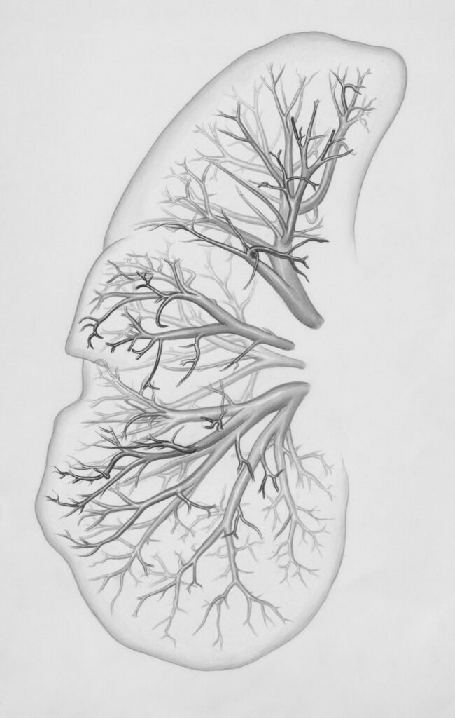
the lower airways, referred to as the tracheobronchial tree, containing (a) the nonrespiratory conducting airways, or the anatomic dead space (i.e., the trachea, bronchi, and bronchioles), that channels inspired air to the gas exchange areas, and (b) the respiratory units, or acini, where gas exchange takes place.
there are 2 lungs right and left which consist on lobes
right lung consists of 3 lobes whereas left lobe consist of 2 lobes
Muscles of respiration
The primary muscles of inspiration are the diaphragm, external intercostal muscles, and parasternal intercostals.
During deep or labored breathing, the accessory muscles of inspiration are recruited. At rest, expiration is a passive process, occurring as the inspiratory muscles relax and lung elastic recoil takes over.
During forced expiration and coughing, the abdominal and internal intercostal muscles are activated.
Respiratory muscle weakness and limited endurance can impair gas exchange and lead to respiratory insufficiency or failure, especially when the mechanics of breathing are altered by hyperinflation of the chest (e.g., emphysema, chronic bronchitis, and acute asthma attack)
blood supply to the respiratory system
The bronchial arteries arising from the descending aorta provide blood supply to the nonrespiratory airways, pleurae, and connective tissue, while the pulmonary arteries supply the respiratory units (acini) and participate in gas exchange.
Numerous pulmonary vasoactive substances can induce vasoconstriction or vasodilation of the pulmonary arterioles.
physiology
the factors that contribute to normal functioning of the respiratory system in order to appreciate normal versus abnormal physiological indicators, both at rest and during exercise, as well as the implications plays vital role in physical therapy interventions
The basic functions of the respiratory system include oxygenation of the blood, removal of carbon dioxide, control of acid–base balance, and production of vocalization.
mechanics of breathing
Respiratory gas exchange requires the movement of sufficient volumes of air into the terminal airways to meet the oxygen needs of the body, whether at rest or during exercise.
This occurs through active contraction of the inspiratory muscles with enough force to override the elastic recoil of the lungs and the resistance to airflow offered by the airways
During Inspiration
during inspiration active muscle contraction results in expansion of the thorax and the lungs, a fall in alveolar pressure, and airflow into the lungs
At rest, inspiration is accomplished primarily by the diaphragm with some assistance from the parasternal and external intercostals and scalenes .
the parasternal intercostals and scalenes act to lift the ribs and expand the upper half of the rib cage, which is important to counteract the inward motion of the upper chest that would result from an unopposed decrease in intrapleural pressure produced by diaphragmatic descent
During exercise the accessory muscles of inspiration are recruited to increase tidal volume (see Table 2-1), which is assisted by passive relaxation of the expiratory muscles that are also activated.
The drop in intrathoracic pressure during inspiration also facilitates venous return to the heart.
During expiration
during expiration passive relaxation of the inspiratory muscles to their resting positions and elastic recoil of the lungs cause alveolar pressure to rise, resulting in airflow out of the lungs.
During exertion, forced expiration, and coughing, active contraction of the expiratory muscles causes a marked rise in intrathoracic pressure so that expiration occurs more rapidly and completely.
passive relaxation of these muscles at end-expiration promotes descent of the diaphragm and induces an increase in lung volume toward its neutral resting position.
references
Frownfelter, D. L., & Dean, E. (2012). Cardiovascular and pulmonary physical therapy: Evidence to practice (5th ed.). Elsevier Health Sciences.
West, J. B. (2012). Respiratory physiology: The essentials (9th ed.). Lippincott Williams & Wilkins.
Moore, K. L., Dalley, A. F., & Agur, A. M. R. (2018). Clinically oriented anatomy (8th ed.). Wolters Kluwer.
Tortora, G. J., & Derrickson, B. (2020). Principles of anatomy and physiology (16th ed.). Wiley.
Palastanga, N., Field, D., & Soames, R. (2012). Anatomy and human movement: Structure and function (6th ed.). Elsevier.
Kisner, C., & Colby, L. A. (2017). Therapeutic exercise: Foundations and techniques (7th ed.). F.A. Davis Company.
Hall, J. E. (2020). Guyton and Hall textbook of medical physiology (14th ed.). Elsevier.
Hough, A. (2017). Hough’s cardio-respiratory care: An evidence-based, problem-solving approach (5th ed.). Elsevier.
Leave a Reply